Recent and Current Research Projects
![]()
Neural Control of Reproductive Function:
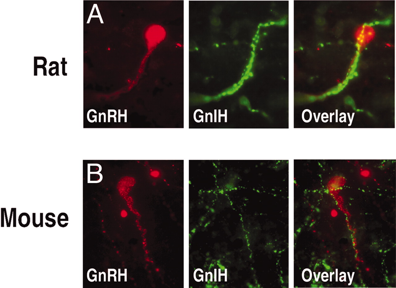
Most generally, my lab is interested in the neural pathways and
neurochemical systems that regulate the reproductive axis. The photomicrograph above shows
projections originating from neurons expressing the RFamide peptide, RFRP-3, a
putatitve gonadotropin-inhibitory hormone (GnIH). We have shown that this peptide inhibits gonadotropin
secretion in rodents, likely via direct projections to the
gonadotropin-releasing hormone (GnRH) systems. [click here to download a pdf of
this manuscript].
![]()
SCN Communication to Central and Peripheral Targets:
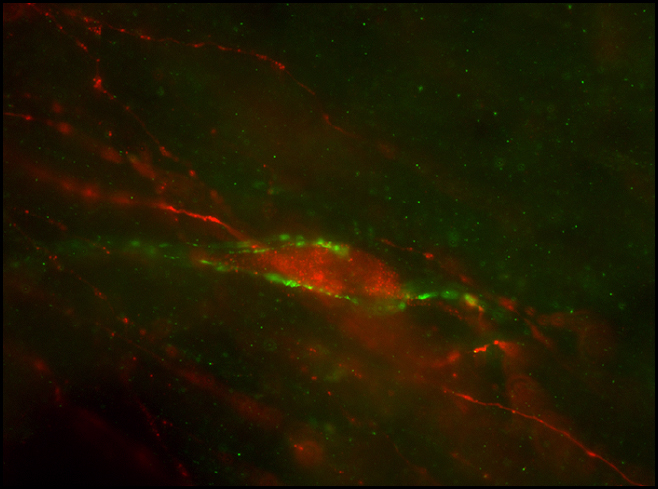
The SCN regulates reproduction by direct projections to the gonadotropin
releasing hormone (GnRH) neuronal system. This photomicrograph shows
projections from vasoactive intestinal polypeptide neurons in the SCN (green)
to a GnRH neuron in the preoptic area (red; a brain area critical for the
regulation of reproduction). [click here to download a pdf of this
manuscript].
![]()
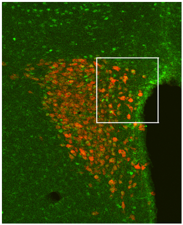
Topographic Organization of SCN Communication:
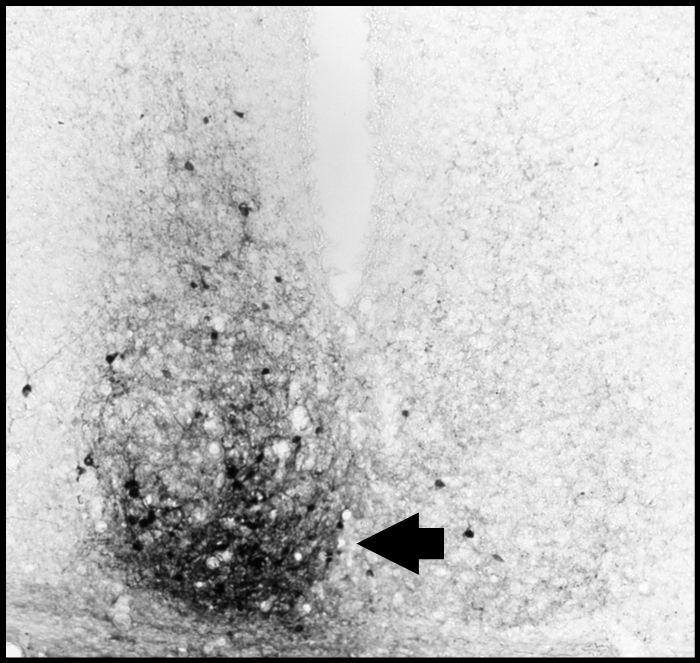
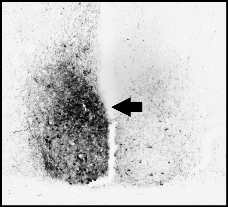
Given the pronounced structural and functional organization of the SCN, it
is important to determine the specific means by which information is
communicated both within the SCN and to SCN targets. In this study,
iontophoretic injections of the anterograde tracer, biotinylated dextran amine
(BDA), were made into subregions of the SCN to determine the specific pattern
of neural communication to all SCN targets.
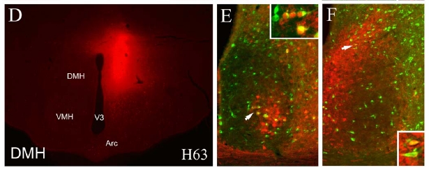
In combination with anterograde injections of BDA, retrograde injections
were made into SCN targets to more specifically determine the SCN cell
phenotypes projecting to effector areas of the brain. (D) Injection of
the retrograde tracer, choleratoxin-beta (CTB), into the dorsomedial
hypothalamus (DMH). (E) SCN of DHM-injected animal stained for both
calbindin (red) and CTB (green). (F) SCN of DHM-injected animal stained
for both vasopressin (red) and CTB (green).
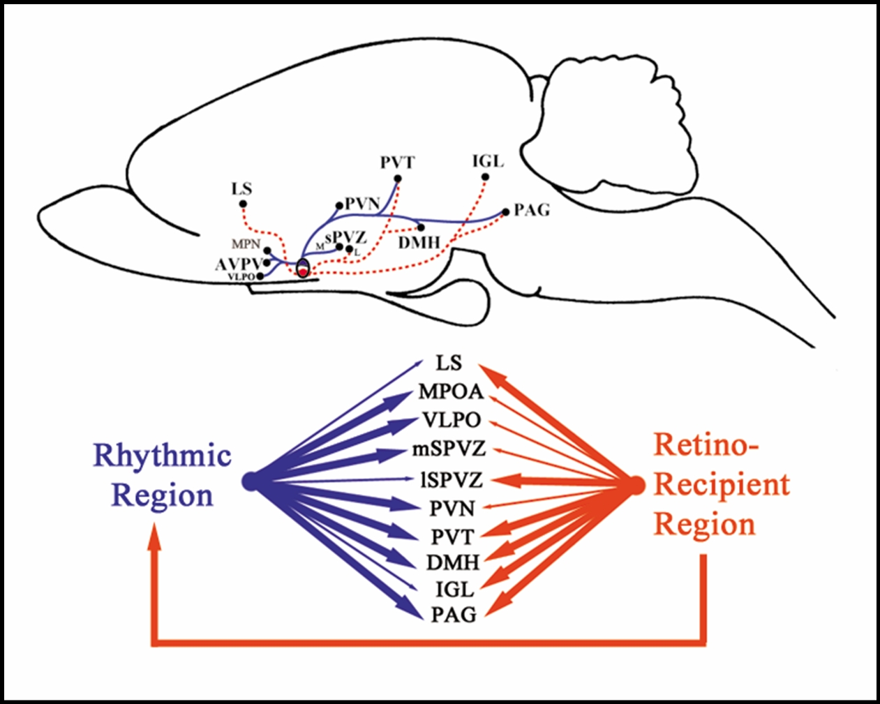
Summary of the topography of SCN communication
revealed by the series of investigations above. Red projections originate
from the calbindin subregion of the SCN while blue projections originate in the
vasopressin cell- and fiber-rich region [click here
to download a pdf of the manuscript].
![]()
Significance of Extra-SCN Clock Genes:

The genes that regulate circadian clock function are not only found in the
SCN, but also in other brain areas and peripheral organs. These
photomicrographs show that neuroendocrine cells in the parventricular nucleus
of the hypothalamus contain clock genes (neuroendocrine cells labeled in red,
the clock gene Period 1 labeled in green). This finding suggests a hierarchical
means by which the SCN regulates the neuroendocrine system, with clock-gene
regulation at various levels of the hypothalamo-pituitary-endocrine gland axis
[click here to download a
pdf of the manuscript].
![]()